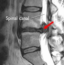
Most of us probably know the situation: The sudden pain at the lower back after a rapid movement or after lifting some weight. Within days this pain starts radiating through buttock and back of the upper leg, even all the way down to the calf. A constant sharp pain that could also wake you up in the middle of the night. And usual pain killing medication like Paracetamol doesn’t work. So you see your doctor or an orthopaedic surgeon who, in many cases, would send you for an MRI scan (magnetic resonance image) of the lower spine straight away. Very often even without a physical exam. This scan will most certainly show a disc prolapse at L5/S1, possibly also at L4/5 level. So there is the apparent ‘reason’ for your pain, and in the worse scenario you will be advised that surgery is the only option to help you.
This diagnosis is unfortunately very often wrong, and the detected disc prolapse is misleading, has nothing to do with the pain and was only found accidentally. Many international studies have shown that up to 80% of people over 40 have a disc prolapse, although they don’t have any symptoms. If we talk about the pre- prolapse stage, the bulging disc, this can be found in 80% of people from the age of 20 onward.
Lower back pain is he most frequent reason for sick leave, and there are many reasons for lower back pain. Most often it would be an inflammation of the sciatic nerve, but also muscular contracture could be responsible for the pain, as well as advanced arthritic changes of the hip joint.
The first step to find the correct diagnosis would be detailed questioning of the patient (anamnesis) about when and how the pain started. Followed by questions about the type of pain – sharp, dull, burning – , how often and exactly where it is felt, how it could be provoked or even released. This would be followed by a thorough physical exam, in which orthopaedic surgeon Dr. Alf Neuhaus always includes exam of the middle spine as well as both hip and knee joints. This could show where the pain comes from, where does is it go to, are neurological symptoms present. In a patient above 40 he would also take radiological images of the pelvis and the lower spine, using the digital X-ray facility available at Clinica SANDALF, to exclude any bony pathologies as possible reason for the pain. The appropriate treatment, which could include physiotherapy, acupuncture, oral medication and even direct infiltration of the inflamed nerve root, could be initiated immediately. In most cases this would already lead to complete relieve of the pain within days. Only if the treatment does not show the desired effect, or neurological symptoms like lack of reflex or significant loss of muscular power, were found during the physical exam, a MRI scan should be performed.
Please don’t hesitate to get in touch with us should you have any further questions regarding disc prolapse, or any other orthopaedic problem.

