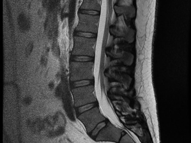
The so called Magnetic Resonance Imaging (MRI) has been first used in medicine for diagnostic findings in the 80th. The area in question is basically screened by a massive magnet. As every tissue is made of atoms with a very specific magnetic ‚spin‘, the MRI can recognize those and construct an image, that can be interpreted by a specialist. And in contrast to radiographic images the MRI scan produces no radiation.
The first step of finding the diagnosis is supposed to be the extensive talk with the patient, to understand and evaluate all details about the symptoms (when did they start, intensity, frequency, quality, previous episodes…). This obviously counts for back and knee pain as well. This so called anamnesis should be follow by a thorough physical exam of the almost naked patient. As an experienced orthopaedic surgeon Dr. Alf Neuhaus would be able to conclude with a very accurate diagnosis, using the information gathered by those two steps. It would also be enough to initiate the appropriate treatment immediately. In case of the patient being over 50, radiographic images should be performed. X-rays of the knees should always be taken in standing position, as it otherwise would be impossible to estimate the degree of arthritic changes. Those radiographic can be performed by orthopaedic surgeon Dr. Alf Neuhaus on the day of your first appointment, using the digital X-ray facilities available in Clinica SANDALF.
Numerous researches have shown that a MRI scan of the lower back or the knees of a person over 50 years of age are not helping to find the diagnosis, as it would show some abnormalities in practically everyone, regardless of any symptoms. MRI scans have technically improved a lot in the last few years, and are now able to show even the smallest anatomical structures. Those detailed images can easily mislead the orthopaedic surgeon to build the wrong diagnosis and to possibly apply an inadequate therapy.
In case of lower back pain a MRI scan only might be needed when all applied treatments like physiotherapy, osteopathy, massage therapy, medication, infiltration and other have been ineffective, or if neurological symptoms like severe numbness, muscle weakness or problems with bladder control, are present.
When it comes to knee problems the step following failed conservative therapy like physio, lymph drainage, infiltration with Cortisone and Hyaluronic Acid would be the joint exploration by key hole surgery (arthroscopy). The arthroscopic evaluation might also be indicated when the knee, following a trauma, has a severe decreased range of movement or is even locked. A special camera is used during this procedure, producing precise pictures of the internal structures of the knee with a 2,5 magnification. Damaged structured, like the meniscus or cruciate ligament, can either be repaired or removed immediately, and cartilage damage can be smoothened. Performing a MRI scan previous to knee arthroscopy does not aid to find the correct diagnosis and does not give any further therapeutic options, and could therefore be considered waste of time and money.
Please get in touch if you would like to talk to us about MRI scan or any other orthopaedic issue.